Back Of Neck Anatomy Muscles : Muscles of the Shoulder and Back Laminated Anatomy Chart ... / This article describes the anatomy of the head and neck of the human body, including the brain, bones, muscles, blood vessels, nerves, glands, nose, mouth, teeth, tongue, and throat.
Back Of Neck Anatomy Muscles : Muscles of the Shoulder and Back Laminated Anatomy Chart ... / This article describes the anatomy of the head and neck of the human body, including the brain, bones, muscles, blood vessels, nerves, glands, nose, mouth, teeth, tongue, and throat.. Intermediate back muscles and c. The head rests on the top part of the vertebral column, with the skull joining at c1. The muscles of the back and neck that move the vertebral column are complex, overlapping, and can be divided into five groups. Muscles make up a large part of the anatomy (structure) of the back. Intermediate layer of back muscles.
There are several individual muscles within the back anatomy, and it's important to take a quick look the image below to shows all the major back muscles (as well as some neck muscles) The three scalene muscles are found forming the floor of the posterior triangle. Intermediate back muscles and c. It's buried under the sternomastoid anteriorly and by. Spinous processes of txi to liii and supraspinous ligaments.
Posterior Neck muscles | Physical Therapy - Powerlifting ... from s-media-cache-ak0.pinimg.com It's buried under the sternomastoid anteriorly and by. They move the head in every direction, pulling the skull and jaw towards the shoulders, spine, and scapula. The muscle is a thick long cord with two heads on the bias coming from the mastoid process through the neck to grudinoklyuchichnomu articulation. Neck muscles help support the cervical spine and contribute to movements of the head, neck, upper back, and posterior longitudinal ligament (pll). The three scalene muscles are found forming the floor of the posterior triangle. Remember that there's a small gap between the clavicles where the manubrium sits, about one eyeball if you're having trouble identifying neck muscles, the levator scapulae is the one that points to the ear. The anterior muscles of the neck facilitate swallowing and speech. Alle muscles are detailed described incl.
The deep back muscles lie immediately adjacent to the vertebral column and ribs.
Some neck muscles attach to the clavicles. There are many muscles around the neck that help to support the cervical spine and allow you to move your head in different directions. In anatomy, the neck is also called by its latin names, cervix or collum, although when used alone, in context, the word cervix more often refers to the uterine cervix, the neck of the uterus.3 thus the adjective cervical may refer. Muscles of the neck are described separately from the compartments. Bones of the neck picture. The neck muscles, including the sternocleidomastoid and the trapezius, are responsible for the gross motor movement in the muscular system of the head and neck. There are several different layers of muscles in your back and often are pulling in different and the intermediate layer of back muscles includes the serratus posterior superior and inferior. The anterior muscles of the neck facilitate swallowing and speech. Remember that there's a small gap between the clavicles where the manubrium sits, about one eyeball if you're having trouble identifying neck muscles, the levator scapulae is the one that points to the ear. Working in pairs on the left and. Several other muscles of the back also extend up to the neck region and are partly connected with the cervical part of the vertebral column, including the trapezius, levator scapulae, splenius, iliocostalis, longissimus, rotatores, semispinalis, interspinales, and intertransversarii muscles. The back anatomy includes the latissimus dorsi, trapezius, erector spinae, rhomboid, and the teres major. There are four pairs of muscles that are responsible for chewing movements or mastication.
This article covers the anatomy of the deep muscles of the back, including their function, blood supply, innervation, origin and insertion. Bodies have two kinds of splenius muscles: The muscles of the back and neck that move the vertebral column are complex, overlapping, and can be divided into five groups. Cervical spine anatomy is quite complex. The anterior and middle scalenes originate from the transverse processes of certain cervical vertebrae and attach to the first rib.
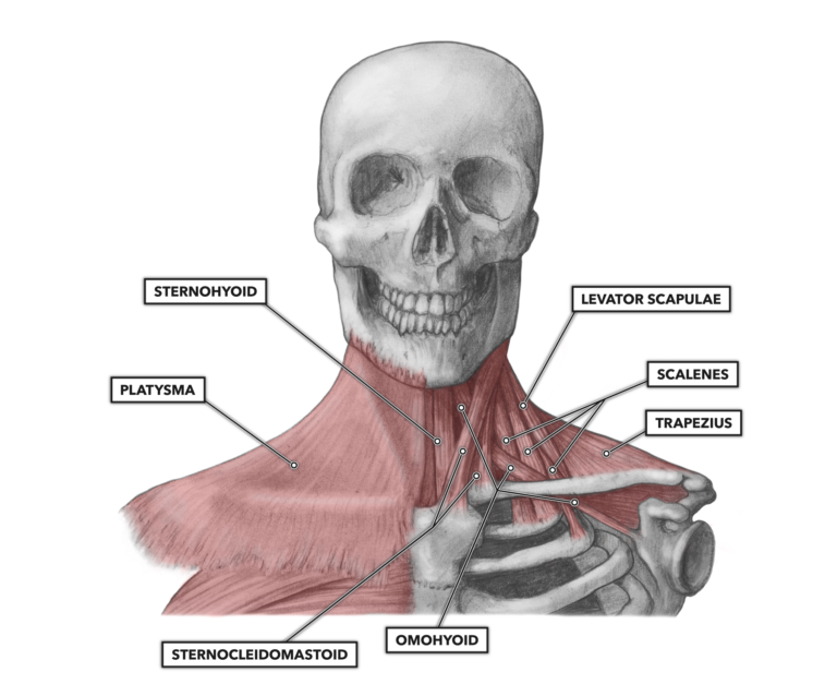
Human neck muscles - Stock Image - F015/8230 - Science ... from media.sciencephoto.com Muscles make up a large part of the anatomy (structure) of the back. Sternohyoid, sternothyroid, thyrohyoid, omohyoid anterior vertebral muscles: The posterior muscles of the neck are primarily concerned with head movements, like extension. Figure 11.13 muscles of the anterior neck the anterior muscles of the neck facilitate swallowing and speech. There are several different layers of muscles in your back and often are pulling in different and the intermediate layer of back muscles includes the serratus posterior superior and inferior. Integrates anatomy and physiology of cells, tissues, organs, the systems of the human body, and mechanisms responsible for homeostasis. The anterior and middle scalenes originate from the transverse processes of certain cervical vertebrae and attach to the first rib. Spinous processes of txi to liii and supraspinous ligaments.
Intermediate back muscles and c.
Spinous processes of txi to liii and supraspinous ligaments. Last update october 2, 2020. Bones of the neck picture. The muscles of the back and neck that move the vertebral column are complex, overlapping, and can be divided into five groups. The neck has no external bone protective structures, so it is quite mobile. There are four pairs of muscles that are responsible for chewing movements or mastication. Sternohyoid, sternothyroid, thyrohyoid, omohyoid anterior vertebral muscles: Several other muscles of the back also extend up to the neck region and are partly connected with the cervical part of the vertebral column, including the trapezius, levator scapulae, splenius, iliocostalis, longissimus, rotatores, semispinalis, interspinales, and intertransversarii muscles. The back anatomy includes the latissimus dorsi, trapezius, erector spinae, rhomboid, and the teres major. We will attempt to provide a simplified overview of this complex anatomy. 12 photos of the muscle anatomy back of neck. Intermediate back muscles and c. In anatomy, the neck is also called by its latin names, cervix or collum, although when used alone, in context, the word cervix more often refers to the uterine cervix, the neck of the uterus.3 thus the adjective cervical may refer.
There are many muscles around the neck that help to support the cervical spine and allow you to move your head in different directions. The neck muscles, including the sternocleidomastoid and the trapezius, are responsible for the gross motor movement in the muscular system of the head and neck. 12 photos of the muscle anatomy back of neck. The back muscles stabilize and move the vertebral. The muscles of the anterior neck are arranged to facilitate swallowing and speech.
CrossFit | Cervical Muscles, Part 1 from www.crossfit.com Bones of the neck picture. This article describes the anatomy of the head and neck of the human body, including the brain, bones, muscles, blood vessels, nerves, glands, nose, mouth, teeth, tongue, and throat. Together they extend neck, and individually they draw and rotate head to one side i.e. Alle muscles are detailed described incl. The back anatomy includes the latissimus dorsi, trapezius, erector spinae, rhomboid, and the teres major. Figure 11.13 muscles of the anterior neck the anterior muscles of the neck facilitate swallowing and speech. The muscles of the back and neck that move the vertebral column are complex, overlapping, and can be divided into five groups. Sternohyoid, sternothyroid, thyrohyoid, omohyoid anterior vertebral muscles:
3d interactive tutorials on the anatomy of the neck, including the anatomical organisation, musculature, larynx, pharynx, blood supply and innervation.
Figure 11.13 muscles of the anterior neck the anterior muscles of the neck facilitate swallowing and speech. Some neck muscles attach to the clavicles. There are several individual muscles within the back anatomy, and it's important to take a quick look the image below to shows all the major back muscles (as well as some neck muscles) The pll starts at c2 and goes down the back of the vertebral bodies and intervertebral discs. They work on the hyoid bone, with the suprahyoid muscles pulling up and the infrahyoid. This article gives an overview of the back's structure and its major muscles. Back pain is common and might be caused by a problem with a muscle. 3d interactive tutorials on the anatomy of the neck, including the anatomical organisation, musculature, larynx, pharynx, blood supply and innervation. The three scalene muscles are found forming the floor of the posterior triangle. The neck muscles, including the sternocleidomastoid and the trapezius, are responsible for the gross motor movement in the muscular system of the head and neck. Muscles of the neck are described separately from the compartments. They are divided into three groups, as shown below. The anterior and middle scalenes originate from the transverse processes of certain cervical vertebrae and attach to the first rib.
Week 2 anatomy (back/neck muscles) back of neck anatomy. Sternohyoid, sternothyroid, thyrohyoid, omohyoid anterior vertebral muscles: